Biology
Part 2: Cell and System Biology
1. Name the basic organelles which make up a eukaryotic cell, and describe their function.
a) Fill in the function of each of the cell structures listed below:
- mitochondria
- rough endoplasmic reticulum
- smooth endoplasmic reticulum
- lysosomes
- nucleus
- nucleolus
- peroxisome
|
- ribosome
- chloroplast
- tonoplast
- centrioles
- Golgi body
- microtubules
|
mitochondria |
generates energy for the cell. |
rough endoplasmic reticulum |
synthesis of proteins destined for export to other membranes or the outside of the cell. |
smooth endoplasmic reticulum |
transport of lipids, proteins, etc. through the cell. |
lysosomes |
intracellular digestion |
nucleus |
storage of genetic material, direction of cell functions |
nucleolus |
synthesis of ribosomal RNA, assembly of ribosomes |
peroxisome |
contains enzymes which transfer hydrogen atoms to oxygen (from various substrates) to form H2O2 which is subsequently broken down. |
ribosome |
protein synthesis |
chloroplast |
photosynthesis: the reduction of CO2 to carbohydrates through the capture of light energy. |
tonoplast |
a large central vacuole in plant cells used to maintain osmotic pressure. |
centrioles |
organization of microtubule assembly during cell division. |
Golgi body |
packaging and preparation of materials for export from the cell. |
microtubules |
support of cell shape, involved in movement of cells, participate in intracellular transport of materials. |
2. Describe the biochemistry of photosynthesis, including light and dark reactions.
a) What do the light reactions supply to the Calvin cycle enzymes?
The light reactions supply ATP and reducing equivalents in the form of NADPH.
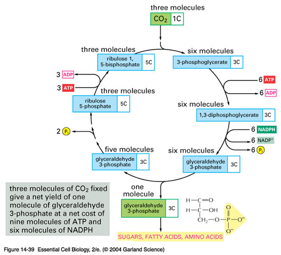
b) What is the overall reaction of the dark reactions?

Note: one inorganic phosphate (Pi) becomes attached to the sugar that is
made, so there are only 17 Pi at the end of the balanced equation.
c) How is oxygen produced during photosynthesis?
Oxygen is produced as a byproduct of the light reacti
ons by photosystem II. The absorption of light at 680 nm by the chlorophyll a molecule allows it to split water into oxygen and hydrogen. The electrons donated from this reaction are carried through various electron carriers to photosystem
I and ultimately to NADPH for use in the dark reactions.
d) How is sugar produced from CO2 during photosynthesis?
Sugar is produced during the dark reactions, also known as the Calvin cycle. Note: to produce one six carbon sugar, there must be two revolutions of the cycle.
e) How many O2 are generated for every CO2 fixed?
One O2 is generated for every CO2 fixed. You need 6CO2 and 12NADPH to generate 1 sugar molecule. This requires splitting 6O2 to generate enough electron reducing equivalents.
3. Name the stages of mitosis, and describe the events which take place at each stage.
a) What are the stages of mitosis?
The stages of mitosis are prophase, metaphase, anaphase, telophase and cytokinesis.
b) What are the major events occurring in each stage?
Prophase: nucleoli and nuclear envelope disappear, chromatin condenses, centriole pairs move away from each other toward the poles. Mitotic spindle forms.
Metaphase: centrioles arrive at the poles, chromosomes bind to the spindle apparatus, and the chromosomes migrate to the metaphase plate, where they align themselves along it, with one chromatid on either side of the metaphase plate and the centromere on the plate.
Anaphase: the centromeres split, and the sister chromatids part. Each sister chromatid moves toward the opposite pole.
Telophase: the chromosomes arrive at the poles, the nuclear membrane and the nucleolus reform, and the chromatin uncoils.
Cytokinesis: a cleavage furrow forms between the daughter cell nuclei, and the cytoplasm divides.
Note: each chromosome prior to anaphase is made up of two chromatids. Once the chromatids separate at anaphase, each is now called a chromosome. Chromatin is the DNA and the proteins which make up the chromosomes.
illustration from: www.accessexcellence.com/AB/GG/mitosis.html
c) Why do drugs which inhibit microtubule polymerization inhibit mitosis?
Inhibition of microtubule polymerization inhibits mitosis because microtubule polymerization is required for formation of the spindle apparatus. If the spindle apparatus doesn't form, then the chromosomes cannot separate and mitosis does not occur.
d) Define diploid.
Diploid refers to any cell which contains two complete sets of chromosomes, one
inherited from each parent during sexual reproduction.
e) What are the functions of the centromere?
The centromere holds the duplicated chromosomes together until they segregate at anaphase. It also provides the binding site for the chromosome to attach to the spindle fibres, and the division point for the separation and movement of the chromosome toward the cell polls during anaphase.
4. Name the stages of meiosis, and describe the events which occur at each stage.
a) What are the stages of meiosis?
Prophase I (leptotene, zygotene, pachytene, diplotene, diakinesis), metaphase I, anaphase I, telophase I, interkinesis, prophase II, metaphase II, anaphase II, telophase II.
b) What are the major events occurring at each stage?
| Prophase I: |
| leptotene: |
chromosomes present as long strands. |
| zygotene: |
homologous chromosomes pair up gene by gene (synapsis). |
| pachytene: |
bivalents (pairs of homologous chromosomes) condense, crossing over occurs. |
| diplotene: |
homologous chromosomes separate in the centromere region, chiasmata (physical attachment at the sites of crossing over) are apparent. |
| diakinesis: |
chiasmata move to the ends of the chromosome arms, nucleolus disappears, nuclear membrane disappears. Spindle apparatus appears. |
|
| Metaphase I: |
chromosomes move to metaphase plate, attach to the spindle apparatus. Once at the metaphase plate, the bivalents orient themselves with one centromere on either side of the plate. |
| Anaphase I: |
Centromeres of the bivalents move toward either pole, the chiasmata move to the ends of the arms, and then disappear. Each homologous chromosome separates and then moves to the opposite pole. |
| Telophase I: |
Chromosomes move to poles, cells divide. Nuclear membrane may or may not reappear at this stage. |
| Interkinesis: |
a short interphase, each cell is 1N at this point, but each chromosome consists of two identical chromatids. |
| Prophase II: |
Like mitotic prophase (nucleoli and nuclear envelope disappear, chromatin condenses, centriole pairs move away from each other toward the poles. Mitotic spindle forms.) |
| Metaphase II: |
Centromeres attach to spindle apparatus, move chromosomes to metaphase plate, centromeres on the plate. |
| Anaphase II: |
Centromeres separate, chromosomes move to poles. |
| Telophase II: |
Chromosomes arrive at poles, nuclear membrane reappears. |
Illustration from: www.accessexcellence.com/AB/GG/meiosis.html
c) How is meiosis different than mitosis?
Meiosis is a reductional division which generates 4 haploid daughter cells from a diploid parent cell. The haploid cells are genetically different from one another. Mitosis conserves chromosomal number, and generates two daughter cells which are genetically identical to each other and to the original parent cell.
d) Assume that an animal has a diploid number of 10. Answer the following questions:
-
How many chromosomes would be present in a typical skin cell?
-
How many chromosomes would be present in a cell at mitotic prophase?
-
How many chromatids would be present in a cell at meiosis I?
-
How many chromatids would be present in a cell at prophase II?
-
How many chromosomes would be present in each gamete?
- Ten chromosomes would be present in a typical skin cell.
- Ten many chromosomes would be present in a cell at mitotic prophase.
- Twenty many chromatids would be present in a cell at meiosis I.
- Ten many chromatids would be present in a cell at prophase II.
- Five many chromosomes would be present in each gamete.
e) Explain the function of crossing-over during meiosis.
Crossing over creates a greater level of genetic diversity - it allows genes that are carried on the same chromosome to be recombined between parental chromosomes to generate new combinations of genes.
illustration from: http://www.accessexcellence.com/AB/GG/comeiosis.html
f) What benefits does crossing-over provide?
Increased genetic diversity.
g) What are chiasmata?
Chiasmata are the physical attachment points which result from cross over events between homologous chromosomes.
5. Describe the process of gametogenesis for male and female animals.
a) What is a polar body?
Polar bodies are the cells which result from the uneven distribution of cellular resources and cytoplasm during oogenesis. Polar bodies remain on the egg surface until they are reabsorbed.
b) Why does gametogenesis in males result in four sperm but in females results in only one egg?
Cell division during spermatogenesis is even - both cells receive half of the available cytoplasm and organelles. Uneven divisions during oogenesis result in
one larger cell with extra growth reserves to support the growth of the zygote. This is necessary to compensate for the fact that sperm contribute little cytoplasm or organelles to the zygote.
c) Meiosis is often described as a reductional division process. Explain this statement.
Meiosis is a reductional division - this means that during cell division, the chromosome number is reduced from 2N to 1N. Thus, when gametes combine, the resulting zygote is diploid once again. (1N + 1N = 2N).
6. Describe how a neuron works and how messages are passed from one neuron to another.
a) What happens when a neuron reaches threshold level?
When a neuron reaches threshold level, an action potential is propagated down the axon.
b) Draw a labelled diagram of the parts of a neuron.
c) Explain how a resting potential develops in neurons.
A resting potential is developed by the action of the Na+ K+ ATPase which helps to maintain the osmotic balance across the cell membrane. This enzyme uses the energy of ATP hydrolysis to pump 3 Na+ out of the cell (to maintain low Na+ levels inside the cell) and 2K+ into the cell (to maintain a high K+ level inside the cell).
The positive charge on the potassium ion helps to balance the high number of positive charges outside the cell from the sodium ions. Note that K
+ can leak freely in and out of the cell due to the presence of leak channels. Eventually a point will be reached at which the electrical forces generated by the attraction of the K
+ ions to the negative charges inside the cell will be balanced by the need of the K
+ ions to move out of the cell down their concentration gradient. The electrical charge needed for this to occur is the resting potential of the neuron.
d) Explain how an action potential propagates down the axon (on the molecular level).
Action potentials propagate down the axon in several steps.
- A stimulus causes a small depolarization of the cell.
- Voltage gated Na+ channels open, allowing Na+ to flow into the cell.
- More voltage gated Na+ channels open in response to the increased depolarization resulting from the Na+ influx. Note: Na+ channels can only stay open briefly - after that they close automatically and are inactive until the resting potential returns. Some neurons can reset more quickly because voltage gated K+ channels open in response to the influx of Na+ (but after a short delay). This decreases membrane depolarization to the resting potential more quickly.
- The depolarization of one point on the axon provides the depolarization stimulus to the next area of the axon and the action potential propagates as a wave down the axon.
e) Why can't an action potential go backwards up the axon?
The action potential can't go backwards up the axon because the Na+ channels are temporarily inactivated after they open until that area of the axon returns the resting potential. Only in the closed state are they able to open in response to a stimulus.
f) What is a neurotransmitter?
A neurotransmitter is a small signaling molecule which is secreted into a synaptic cleft that serves to modify the activity of the post synaptic neuron and pass a signal between the two neurons.
g) How does an action potential cause the release of neurotransmitters?
When the action potential reaches the end of the axon, it causes voltage gated Ca2+ channels to open in response to the membrane depolarization which is occurring. This allows an influx of Ca2+ into the cell (Note: cells normally maintain an internal concentration of Ca2+ that is 1000 times lower than the external environment). The influx of Ca2+ causes secretory vesicles containing neurotransmitter to fuse with the presynaptic membrane and release the neurotransmitter into the synaptic cleft.
h) Explain in detail how a message moves from one neuron to the next.
- The action potential depolarizes the membrane.
- Voltage gated Ca2+ channels open.
- Secretory vesicles fuse with presynaptic membrane in response to Ca2+ influx.
- Neurotransmitter is released and diffuses across the synaptic cleft.
- If the synapse is inhibitory:
- transmitter gated chloride channels open and the production of action potentials is inhibited.
If the synapse is excitatory:
- transmitted gated Na+ channels open.
- membrane becomes slightly depolarized.
- if enough channels open, an action potential results
7. Describe antigen-antibody reactions.
a) What is an antigen?
An antigen is any molecule capable of generating an immune response. Most are greater than 10000 daltons in molecular weight (1 dalton = molecular weight of hydrogen), and they are usually either proteins or polysaccharides.
b) What portion of an antibody does an antigen bind to?
An antigen binds to the antigen binding sites at the ends of the arms of the antibody.
c) What is the role of the Fc portion of the antibody?
The Fc portion of the antibody binds to immune cells - it can be used to anchor antibodies to the surface of B cells, and may also help antigens coated with antibodies to be taken up by macrophages.
8. Describe the function of cellular and humoral immunity.
a) What is the difference between humoral and cell mediated immunity?
Immunity is humoral if a soluble antigen interacts directly with antibodies. B cells amplify the antibody response. Cell mediated immunity is when the antigens are intracellular, and the immune response depends on the production and amplification of T cells.
b) What is an antigen presenting cell?
A cell which engulfs an antigen and displays its digested parts on the cell surface. The antigen presenting cell (APC) can be contacted by a T cell, which may become activated in response to the antigen fragments being "presented" in the context of other cell surface markers.
c) What is a cytokine?
A cytokine is a soluble immune response modulator produced by leukocytes (white blood cells). In response to cytokine production, immune cells may be attracted to the area, proliferate, etc. depending on the situation.
d) Describe how the theory of clonal selection relates to antibody production.
The theory of clonal selection relates to antibody production in that rather than always having large amounts of every possible antibody around or large numbers of cells that could make every possible antibody, the body has a few B cells of every type around. These B cells do not reproduce in the absence of the appropriate antigen. The antigen then "selects' the correct B cell clones to amplify - those cells whose surface antibody binds to the antigen. These cells can then generate a large population of effector cells which secrete large amounts of the specific type of antibody needed.
e) A person is infected with a virus (an intracellular pathogen). Outline the steps the immune response will use to eliminate the virus.
Intracellular pathogens require a cell mediated immune response. An antigen presenting cell such as a macrophage will engulf the antigen and display digested parts of it on the cell surface in combination with the MHCII protein complex (Major Histocompatibility Complex). The MHCII protein complex is a cell surface marker for immune cells.
The activated macrophage will bind to T cells with receptors that recognize the antigen fragments, causing the macrophage to secrete the cytokine IL-1 (interleukin 1). This will cause T helper (TH)and T cytotoxic (Tcyt)cells to proliferate. The Tcyt cells will recognize infected cells by looking for pieces of virus on the surface of infected cells in combination with MHCI (a protein complex present on the surface of all nucleated cells in the body which allows the immune system to recognize them as "self"). When Tcyt cells bind to infected cells they release perforin, a protein complex which causes the infected cells to lyse.
f) A person is infected with Staphylococcus sp. (an extracellular pathogen). Outline the steps necessary for the immune response to eliminate the infection.
An extracellular pathogen requires a humoral response. The antigen will enter the blood, coming in contact with a B cell which has antibodies on its surface which match the antigen. The B cell will grow and multiply in response to contact with the antigen, and most of the progeny cells will differentiate into plasma cells (antibody secreting cells which can secrete up to 2000 antibodies per second for their 4 - 5 d lifespan).
The antibodies produced by the plasma cells will tag the antigen (form antigen-antibody complexes). At this point, the complexes may be phagocytosed by macrophages, may cause agglutination (formation of very large, crosslinked complexes) which may be removed by the kidneys or phagocytosed, or the antigen-antibody complexes may activate the complement cascade, which will result in lysis of the antigen cells.
g) Explain how the skin can be considered to be part of the immune system.
The skin can be considered to be part of the immune system because it represents a physical barrier to the microbial colonization of the interior of the body. It is a comparatively dry, salty, low pH environment due to the products of the sebaceous (sweat) glands. In addition, the normal flora of the skin (those bacteria, etc. that are normally present on healthy human skin) will compete with pathogens for resources such as space and nutrients.
9. Differentiate between peptide and steroid hormones.
a) Describe the structure of peptide and steroid hormones.
Steroid hormones are fat soluble molecules whose structure is based on
that of cholesterol. Peptide hormones are short chains of amino acids -
they may be as short as 3 amino acids or as long as 200 amino acids.
b) How are peptide and steroid hormones different in terms of their cellular activity?
Steroid hormones pass through the cell membrane of target cells by passive diffusion, and they bind to protein receptors in the nucleus. The hormone-receptor complex binds to a specific site on the DNA (the hormone response element) and alters gene expression.
Peptide hormones bind to protein receptors on the cell surface. The
receptor interacts with a second messenger system. The second messenger
produces a signal that elicits a cellular response, from proteins that
already exist in the cell.
10. Discuss how hormones work, and the role of various endocrine organs in the functioning of a healthy body.
a) What is a hormone?
A hormone is a molecule synthesized and secreted by an endocrine gland into the blood which travels to a distant site and elicits a specific biological response from the target cell.
b) What are some of the important functions of hormones?
Functions of hormones include: sexual differentiation; regulation of blood sugar levels, Ca2+ levels, growth, milk production, blood osmolarity, metabolic rate, etc.
c) Describe the actions of prolactin, thyroid stimulating hormone, estrogen, and growth hormone.
- prolactin: stimulation of milk production and secretion in humans.
- thyroid stimulating hormone: stimulates the thyroid gland to secrete hormones such as T3 (triiodothyronine) and T4 (thyroxine) which govern metabolic rate.
- estrogen: initiates building of the uterine lining, develops and maintains female sexual characteristics.
- growth hormone: stimulates general growth, especially of skeleton, and affects metabolism.
d) What two types of hormones are released by the adrenal cortex and what are the actions of each?
- glucocorticoids: increase blood sugar by altering many aspects of carbohydrate metabolism.
- mineralocorticoids: promote the readsorption of Na+ and excretion of K+ by the kidneys.
e) What are prostaglandins? What do they do?
Prostaglandins are modified fatty acids which act as local regulators. E.g. they may be involved in relaxing/contracting blood vessels (PG E/PG F), or in uterine contractions during labour.
f) Explain the role of cAMP in hormone action.
Cyclic AMP (cAMP) is a second messenger for hormone action. When the hormone binds to its receptor, the receptor activates the enzyme adenylate cyclase which converts ATP to cAMP and pyrophosphate. The rise in internal cAMP levels causes the cellular response, often in the form of an enzyme cascade. Because cAMP has a short half-life in the cell, it breaks down quickly and allows this system to respond rapidly to changes in the levels of hormone acting at the receptor.
cAMP may also be generated indirectly - the receptor may interact with a G protein and the G protein can then interact with adenylate cyclase.
g) Explain how insulin interacts with its target cells.
Insulin has a receptor made up of α chains and β chains. The insulin binds to the α chains, which causes the activation of the tyrosine kinase of the β chains (which autophosphorylate themselves). This causes other enzymes to phosphorylate other proteins which result in:
a) increased uptake of glucose by cells (liver and muscle).
b) increased synthesis, and decreased breakdown, of glycogen.
c) increase in glycolysis activity
d) increased fatty acid and triacylglycerol synthesis.
h) Explain the role of the pituitary gland.
The pituitary gland has two functional parts:
- the anterior pituitary which responds to signals from hypothalmic hormones to release ACTH, TSH, FSH, LH, GH, etc.
- the posterior pituitary which contains the axon termini of many hypothalmic neurons. This area also secretes hormones including oxytocin, vasopressin and ADH.
The pituitary mediates signals from the brain to the rest of the endocrine system.
i) Draw a zinc finger from a steroid hormone receptor.
j) Explain how zinc finger proteins activate genes with their hormone partner.
Zinc finger proteins have 3 distinct regions - a transcription activation sequence, a DNA binding element and a hormone binding element.
The DNA binding element is the zinc finger region. The ends of the "fingers" interact with the bases in the DNA by inserting themselves into the major groove of the double helix of DNA. Following the binding of the hormone-receptor complex to the hormone response element, the transcription activation domain alters the expression levels of the adjacent genes.